Great geoscience photographs aren’t always shots of beautiful landscapes. Sometimes there are stunning things to see at a much smaller scale. This week’s Imaggeo on Mondays showcases one such curiosity and highlights how research images can reveal a lot about the natural world when exhibited as a form of art.
Thin sections are a fantastic way of finding out more about rocks, soils and tissue structure. At 30 micrometres thick they are the most refined Carpaccio you could find on a geoscientist’s plate, and illuminating them under a microscope only makes the sections more splendid.
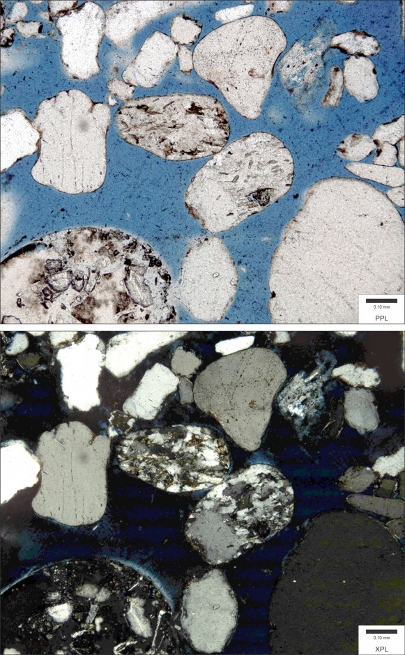
A sandstone sample viewed in plane polarised light (top) and cross polarised light (bottom). (Credit: Wikimedia Commons user Michael C. Rygel)
Under plane polarised light, you spot the fine details that make up each slice, the crystal grains, pore spaces and shell fragments that, together, make up your rock sample. And in cross polarised light there’s even more to be seen. Crystals, at first barely perceptible, shine out amid dark masses and appear as an array of bright and beautiful colours. Different properties like the refractive index and extinction of a crystal can let you work out what mineral you’re looking at, and the relationships between mineral grains offer clues to the rock’s history.
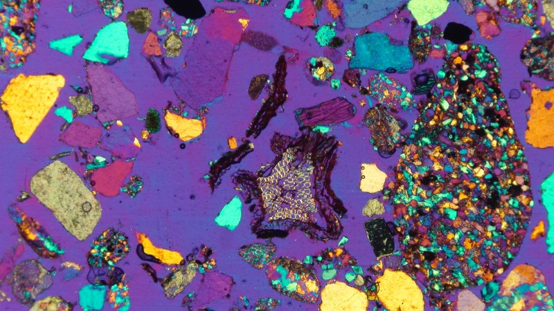
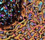
Under the microscope, where mineral and biological worlds meet. (Credit: Laura Gargiulo via imaggeo.egu.eu)
This image, by Laura Gargiulo, shows the surface of a sandy soil under cross polarised light. Sand grains – a veritable pick a mix of rock fragments and merged minerals – make up the majority and a slice of cellular plant material sits just south of the centre. Each of the colours and the way they change under polarised light reveal what each sand grain is made of and how these tiny fragments combine to make up the soil. While its purpose may be a scientific one, the image certainly has aesthetic appeal.
By Sara Mynott, EGU Communications Officer
If you are pre-registered for the 2014 General Assembly (Vienna, 27 April – 2 May), you can take part in our annual photo competition! Up until 1 March, every participant pre-registered for the General Assembly can submit up three original photos and one moving image related to the Earth, planetary, and space sciences in competition for free registration to next year’s General Assembly! These can include fantastic field photos, a stunning shot of your favourite thin section, what you’ve captured out on holiday or under the electron microscope – if it’s geoscientific, it fits the bill. Find out more about how to take part at http://imaggeo.egu.eu/photo-contest/information/.